This activity is provided by Med Learning Group.
This activity is supported by an educational grant from Regeneron Pharmaceuticals, Inc.
Copyright © 2019Med Learning Group. Built by Divigner. All Rights Reserved.
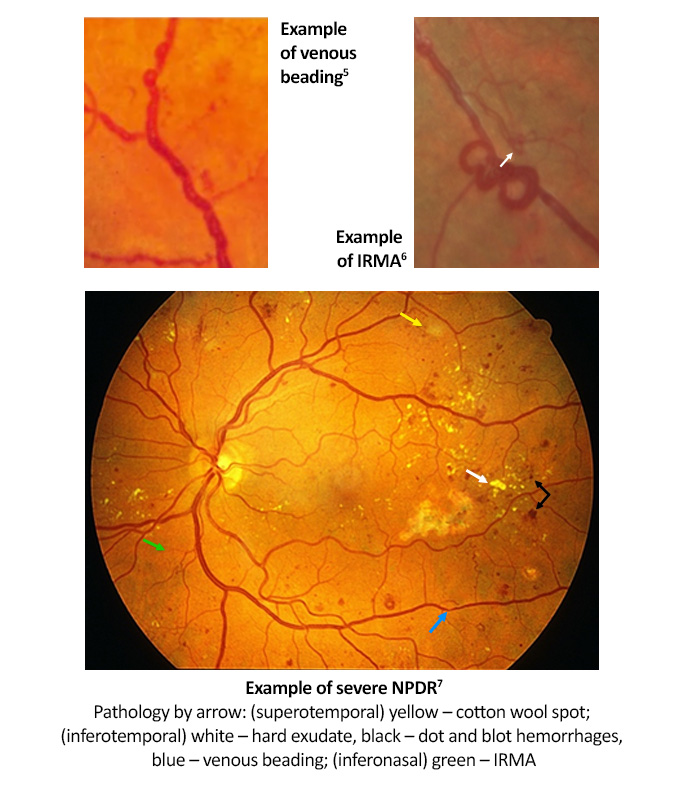
Diabetic retinopathy (DR) is the most common cause of vision loss in working-age adults, affecting one in three people with diabetes.1,2 Early recognition allows for timely treatment and improved visual prognosis.1,2 Many people with nonproliferative diabetic retinopathy (NPDR) do not experience symptoms until the retinopathy is severe.1
However, despite a person with diabetes having good vision, they may still have significant retinal disease, making regular eye screenings essential.1 Unfortunately, only around 60% of people with diabetes have recommended yearly screenings for diabetic retinopathy.1
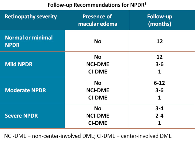
Diagnosis of NPDR involves a complete eye exam, including dilated fundus exam; only 50% of eyes are correctly diagnosed through undilated pupils.1 Care should be taken to examine the mid-peripheral and peripheral retina as an estimated 30% of people with NPDR have retinal abnormalities outside the posterior pole.4 Additional testing with optical coherence tomography (OCT) or fluorescein angiography (FA) may be indicated, especially if there is a concern for diabetic macular edema (DME).3 The follow-up schedule depends on the severity of NPDR and the presence of DME, along with individual patient factors.1
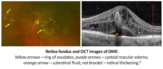
DME can occur at any stage of DR but is more commonly noted with increasing retinopathy severity.8 Reports of DME incidence are as high as 25% to 29% among people with type 2 and type 1 diabetes, with overall estimated US prevalence in persons with diabetes anywhere from 2.7% to 3.8%; if left untreated, DME can lead to significant vision loss.8
Recent guidelines classify DME into two types: center-involved and non-center-involved DME.2 Center-involved DME demonstrates retinal thickening within the 1 mm diameter central subfield, whereas non-center involved DME does not involve this area.2 Optical coherence tomography (OCT) findings include retinal swelling, cystoid macular edema, and subretinal fluid.8
Associated comorbidities with NPDR include hypertension and hyperlipidemia; either condition increases the risk of developing DR.1,2 People with metabolic syndrome (a collection of risk factors that increase the risk of developing heart disease, diabetes, and stroke) have a higher risk of DR progression.1
All URL’s accessed 5/9/22.
This activity is provided by Med Learning Group.
This activity is supported by an educational grant from Regeneron Pharmaceuticals, Inc.
Copyright © 2019Med Learning Group. Built by Divigner. All Rights Reserved.
James P. Gills Professor of Ophthalmology
Professor of Ophthalmology, Johns Hopkins University School of Medicine
Wilmer Eye Institute, Johns Hopkins Medicine
Baltimore, MD
Specializing in Diabetes Eye Care & Education, Chous Eye Care Associates
Adjunct Professor of Optometry, Western University of Health Sciences
AOA Representative, National Diabetes Education Program
Tacoma, WA
Chief of Optometry, Sepulveda VA Medical Center
Professor, Southern California College of Optometry at Marshall B. Ketchum University
Sepulveda, CA
Ophthalmologist-in-Chief
Wills Eye Hospital
Philadelphia, PA
Director, Retina Research
Wills Eye Hospital
Professor and Chair of the Department of Ophthalmology
Thomas Jefferson University Hospitals
Philadelphia, PA
Director of Research, Retina Consultants of Houston
Associate Professor of Clinical Ophthalmology
Blanton Eye Institute & Houston Methodist Hospital
Houston, TX
The RELIEF Clinical Toolkit is an online tool that aims to provide clinicians with up-to-date information on the presentation, prognosis, pathophysiology, and treatment strategies for diabetic retinopathy (DR) in patients with diabetes who have or are at risk for developing DR. Click on one of the options below to learn more about DR.
This activity is provided by Med Learning Group.
This activity is supported by an educational grant from Regeneron Pharmaceuticals, Inc.